Monday 31 August 2015

Replication of positive sense RNA Virus


With the exception of retroviruses and some unusual viruses related to viroids, single-stranded (ss) RNA virus genome replication requires two stages. First, the input strand must be transcribed (using Watson-Crick base-pairing rules) into a strand of complementary sequence and opposite polarity. Replication occurs as a “fuzzy,” multibranched structure. This complex, dynamic structure contains molecules of viral transcriptase (replicase), a number of partially synthesized product RNA strands (“nascent” strands), and the genome-sense template strand. The whole ribonucleoprotein (RNP) complex is termed the type 1 replicative intermediate or RI-1. The single-stranded products generated from RI-1 are antisense to the genomic RNA. This complementary strand RNA serves as a template for the formation of more genomic-sense RNA strands. This second replicative intermediate (RI-2) is essentially the same in structure as RI-1 except that the template strand is of opposite sense to genomic RNA and the nascent product RNA molecules are of genome sense.

Remember that (1) Virion RNA is the template in RI-1. (2) RI-1 produces template RNA of opposite sense to virion RNA. (3) RNA that is complementary to virion RNA is the template in RI-2. (4) RI-2 is the intermediate for expression of RNA of the same sense as the virion.

One further general feature of the replication of RNA viruses is worth noting. The error frequency (i.e., the frequency of incorporating an incorrect base) of RNA-directed RNA replication is quite high compared to that for dsDNA replication. Thus, typically DNA-directed DNA replication leads to incorporation of one mismatched base per 107 to 109 base pairs, while RNA directed RNA synthesis typically results in one error per 105 bases. Indeed, the error rate in the replication of some RNA genomes can be as high as one error per 104 nucleotides.

Part of the reason for this error rate for RNA is that there is no truly double-stranded intermediate; therefore, there is no template for error correction or “proofreading” of the newly synthesized strand as there is in DNA replication. A second reason is that RNA polymerases using RNA templates seem to have an inherently higher error frequency than those utilizing DNA as a template. For these reasons, infection of cells with many RNA viruses is characterized by the generation of a large number of progeny virions bearing a few or a large number of genetic differences from their parents. This high rate of mutation can have a significant role in viral pathogenesis and evolution; further, it provides the mechanistic basis for the generation of defective virus particles. Indeed, many RNA viruses are so genetically plastic that the term quasi-species swarm is applied to virus stocks generated from a single infectious event, as any particular isolate will be, potentially at least, genetically significantly different from the parental virus.


 Some general features of viruses containing RNA genomes that use RNA-directed RNA transcription in their replication. a. The general relationship between viruses containing a genome that can be translated as the first step in the expression of viral genes versus those viruses that first must carry out transcription of their genome into mRNA utilizing a virion associated transcriptase. b. The basic rules for RNA-directed RNA replication. As with DNA-directed RNA and DNA synthesis, the new (nascent) strand is synthesized 5′ to 3′antiparallel to the template, and the Watson-Crick base-pairing rules are the same, with U substituting for T. However, the very high thermal stability of dsRNA leads to complications. The major complication is that newly synthesized RNA must be denatured and removed from the template strand to avoid its “collapsing” into a double-stranded form. Formation of such dsRNA is an effective inducer of interferon, and it appears to be refractory to serving as a template when free in the cytoplasm. A second complication is that in order to generate an ssRNA molecule of the same coding sense as the virion genome, two replicative intermediates (RI’s) must be generated. These intermediates are dynamic structures of ribonucleoprotein containing a full-length template strand, and a number of newly synthesized product RNA molecules growing from virion encoded replicase that is traversing the template strand. RI-1 generates RNA complementary to the virion genomic RNA. This serves as a template for new virion genome RNA in RI-2.


Saturday 29 August 2015

Cap- and poly(A)-independent initiation in translation in Viruses


Several groups of positive-strand RNA viruses containRNAs that are neither capped nor polyadenylated. These viruses have evolved alternative strategies for translation that use 3′-UTR enhancer elements to recruit eIF4F or eIFiso4F and/or to base pair with their 5′-UTRs for loading
the 43S preinitiation complex at the 5′- start site. Satellite tobacco necrosis virus (STNV – a positive strand RNA necrovirus) RNA contains within its 3′-UTR (just after the termination codon of the coat protein coding region) a translational enhancer domain (TED) that promotes efficient cap-independent translation when combined with the STNV 5′-UTR (for STNV-1 strain see Timmer etal. 1993). Although TED can functionally substitute for a 5′-cap structure, its function in vitro is dependent on the presence of eIF4F and eIFiso4F (Timmer et al. 1993). Indeed TED specifically binds eIF4E and eIF4iso4Ein vitro (van Lipzig et al. 2002; Gazo et al. 2004), while the 5′-UTR of STNV-1 has the potential to base pair with TED and the 3′-end of 18S rRNA (Timmer et al. 1993).

Thus to promote cap-independent translation initiation, TED recruits the 43S preinitiation complex by binding canonical cap-binding factors at the 3′-UTR, while potential base pairing between the viral 5′- and 3′-UTRs would be required for transfer of this 43S complex to the 5′-UTR to locate the initiation codon. The existence of the 3′- to 5′-UTR pathway to recruit the translational machinery is probably not unique to STNV TED, but is likely to apply to other enhancer elements present within the 3′-UTRs of other non-capped, non-polyadenylated virus RNAs. Barley yellow dwarf virus (BYDV – a luteovirus) RNA also lacks a 5′-cap structure and poly (A) tail, but it harbors a cap-independent BYDV translational element (BTE) functionally similar to TED within the 3′-UTR (Wang et al. 1997; Guo et al. 2000). A BYDV like BTE is present in all Luteoviruses, as well as in Dianthovirus [Red clover necrotic mosaic virus (RCNMV), Mizumoto et al. 2003] and Necrovirus [tobacco necrosis virus (TNV), Meulewaeter et al. 2004; Shen and Miller 2004], and contains the conserved sequence CGGAUCCUGGGAAACAGG, which also functions when placed in the 5′-UTR. In its natural location this sequence has the potential to base pair to the 5′-UTR (Wang et al. 1997; Guo et al. 2000, 2001). BTE can recruit the translation machinery to the 3′-end and deliver it to the 5′-UTR by a 3′–5′ RNA interaction (Wang et al. 1997).

The delivery of the translational machinery to the 5′-end can occur due to long-distance kissing-loop interactions between RNA hairpins in BTE and the 5′-UTR (Guo et al. 2001). Thus, TED and BTE behave in a similar way to strongly stimulate cap-independent translation, without exhibiting any conservation of sequence or secondary structure. Whether BTE or TED require participation of canonical translation initiation factors for their action remains to be investigated. Another distinct enhancer element identified in Tombusvirus, Tomato bushy stunt virus (TBSV), has been termed the 3′-cap-independent translational enhancer. The TBSV 5′-UTR folds into a complex RNA structure, which was recently demonstrated to physically interact with the 3′CITE in vitro (Fabian and White 2004). Formation of 5′–3′ RNA interactions correlates well with efficient translation in vivo and might support the transfer of the translational machinery from the 3′ to the 5′-end of the RNA as suggested for BTE and TED (Figure 5).


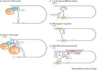
 3′ cap-independent translational enhancer (3′ CITE)-mediated translation. a | General 3′ CITE model showing the long-range interaction (indicated by the double-headed arrow) that positions the eukaryotic translation initiation factor 4F (eIF4F)-bound 3′ CITE close to the 5′ end of the genome, where it enables eIF4F-mediated recruitment of the 40S ribosomal subunit to the 5′ end to initiate translation. Interacting sequences are shown in green. b | Dual 3′ CITE model. Pea enation mosaic virus (PEMV) contains two different types of 3′ CITE: a panicum mosaic virus-like translational enhancer (PTE), which is located in the 3′UTR and binds to eIF4F; and a kissing-loop T-shaped structure (kl-TSS), which is positioned immediately upstream and binds directly to the 60S ribosome subunit and mediates long-distance RNA–RNA base pairing with a 5′-proximal hairpin. c–e | Internal ribosome entry site (IRES)-mediated translation. c | The foot-and-mouth disease virus (FMDV) IRES is stimulated by the 3′ UTR, which engages in long-range contacts with two regions in the 5′ UTR: the IRES and a region that has been shown to be involved in genome replication, which is known as the S-region. The specific sequences that are involved in this interaction have not been identified and the interaction with the S-region may modulate genome replication. d | The 3′-terminal hexamer CGGCCC in classical swine fever virus (CSFV) is a negative modulator of IRES-mediated translation and may confer its inhibition by pairing with a ribosome-binding region in the IRES, thus blocking ribosome binding. e | Hepatitis C virus (HCV) IRES activity is negatively regulated by an interaction between helix IIId of the IRES and a bulge in the structure 5BSL3.2, which is located in the coding region of non-structural protein 5B (NS5B). The same bulge in 5BSL3.2 also interacts with a genomic sequence around position 9110 of the genome, and the terminal loop of 5BSL3.2 can pair with the 3′ SL2 element located in the 3′ UTR, which may modulate genome replication. These interactions may coordinate viral translation and genome replication.

Thursday 27 August 2015

Cap-independent translation of proteins in Viruses


Some (+) ssRNA plant viruses, including those of the large families Potyviridae and Comoviridae, have replaced the cap by a small protein, VPg (Viral Protein genome-linked), which is covalently attached to the first nucleotide of the mRNA. Potyviruses and comoviruses closely resemble animal picornaviruses and caliciviruses in genome structure and in having a VPg. However, all these viruses possess a conventional poly(A) tail. Lacking a cap, they seem to rely on an internal initiation pathway of translation.

The internal ribosome entry site (IRES) is an RNA sequence that promotes direct binding of the 40S ribosomal subunit to internal regions of mRNA, usually upstream of the major ORF start site, thus skipping the requirement for the cap-binding protein, eIF4E (Hellen and Sarnow 2001). The IRESes of animal picornaviruses were the first to be discovered and remain the most extensively studied RNA elements that drive internal initiation. The RNA genomes of picornaviruses contain a very long structured leader with an IRES that promotes entry of the 40S ribosomal subunit near the AUG start codon for the major polyprotein. Direct binding of eIF4G to the encephalomyocarditis virus (EMCV) IRES is required for 40S ribosomal subunit entry via IRES/eIF4G/ eIF3/40S interactions (Pestova et al. 1996a, 1996b). A similar mechanism appears to operate in plants for at least two representatives of picorna-like potyviruses: tobacco etch virus (TEV, Carrington and Freed 1990) and turnipmosaic virus (TuMV, Basso et al. 1994).

The 5′-UTRs of TuMV and TEV differ from those of animal picornaviruses by their smaller length and less complex secondary structure. Zeenko and Gallie (2005) demonstrated that a pseudoknot element (PK1) of the TEV leader is a core structure of the IRES and is sufficient to promote cap-independent translation. However, under these conditions translation still required eIF4G (Gallie 2001). Binding of the 40S ribosome to the IRES is apparently mediated by eIF4F and eIFiso4F, since direct binding of eIF4G and eIFiso4G, as well as their complexes (eIF4F and eIFiso4F), to the TEV 143-nt 5′-leaderand PK1 was recently demonstrated, whereby eIF4G (or eIF4F) possesses stronger affinity to the TEV leader than does eIFiso4G (Ray et al. 2006).

The IRES preferentially recruits eIFiso4F to form the 48S preinitiation complex. At the same time, it is puzzling that the 5′-end of TEV genomic RNA can recruit eIFiso4E via VPg protein and that this interaction is strong enough to compete with cap binding in Arabidopsis (Miyoshi et al. 2006) despite the fact that VPg and cap bind to different sites of eIF4E (Léonard et al. 2000). Corresponding interactions between eIF4G, eIF4E and VPg have recently been demonstrated for lettuce mosaic virus (LMV; Michon et al. 2006) and TuMV (Miyoshi et al. 2006). Although the role of eIF4G/eIF4E/VPg complexes in regulation of translation initiation is not yet clear, their participation in IRES-mediated translation initiation has been proposed for TuMV (Khan et al. 2006). Indeed, in mammalian positive-stranded RNA caliciviruses, translation initiation is strictly dependent on an interaction between VPg and eIF4E, where VPg seems to substitute for the cap (Goodfellow et al. 2005). Whether VPg can substitute for cap in plants still needs to be clarified. The fact that PABP was found to be required for full IRES function invitro (Gallie 2001) suggested that TEV genomic RNA can be circularized via eIF4G, eIF4iso4G and PABP. The RNA polymerase (RdRp) of Zucchini yellow mosaic virus (ZYMV; Wang et al. 2000) and VPg-Pro of TuMV (Léonard et al. 2004) interact with PABP and can achieve circularization between 5′- and 3′- termini to facilitate replication. Although shutdown of the host translation machinery by plant viruses was not demonstrated, increasing concentrations of RdRp might interfere with the translation process.

VPg can recruit eIF4E (or eIFiso4E) for the benefit of the virus in different host plants: eIFiso4E recruitment by TEV VPg is required to infect Arabidopsis plants, while eIF4E recruitment is required for infection of solanaceous species (for a review, see Robaglia and Caranta 2006). The respective resistance genes of naturally occurring potyvirus resistant plant varieties have been shown to encode defective eIF(iso)4E or eIF4E (Lellis et al. 2002; Ruffel et al. 2002). Interestingly, involvement of a VPg/eIFiso4E complex in cell-to-cell movement or viral transport has also been suggested (Dunoyer et al. 2004; Gao et al. 2004).

Putative, unstructured IRESes of tobamoviruses

IRESes identified in plant tobamoviruses are unusual in that they are very short and unstructured. The RNA genome of tobamoviruses, of which tobacco mosaic virus (TMV) strain U1 is the type member, is a polycistronic capped RNA containing four large ORFs. The first ORF and its read-through ORF, which encode the two components of viral replicase, are translated directly from the genomic RNA. The virus produces two subgenomic (sg)RNAs: the dicistronic I2 RNA is used for translation of movement protein (MP), and a monocistronic RNA codes for CP (Palukaitis and Zaitlin 1986). Another member of the tobamoviruses, a crucifer-infecting virus (crTMV; Dorokhov et al. 1994), has a similar organization but its MP and CP ORFs overlap. crTMV harbours two unusual IRESes, the 148 nt  and the 75 nt IRESMP, which are thought to drive translation of the CP (Ivanov et al. 1997) and MP (Skulachev et al. 1999) ORFs, respectively. Both contain a purine- rich tract upstream of the AUG start codon. Interestingly, IRESCP can function in plants, yeast and HeLa cells (Dorokhov et al. 2002). However, the functional significance of these IRESes is obscure since both MP and CP canbe also translated from sgRNAs by a ribosome scanning mechanism (Ivanov et al. 1997; Skulachev et al. 1999). Although the IRESCP region is highly conserved between crucifer- infecting tobamoviruses, it differs from other tobamoviruses (Ivanov et al. 1997). Another very short purine-rich IRES element was identified upstream of the MP ORF of TMV U1 (Skulachev et al. 1999), which could be preferentially used for TMV U1 I2 RNA due to the possible lack of a 5′ cap in this RNA. The short purine-rich stretch was suggested as an IRES module that can mediate internal initiation in plants, mammals and yeast (Dorokhov et al. 2002).

Poly(A)-independent initiation

The positive-strand RNA genomes of Bromoviridae and Tobamoviridae have a 5′-cap and a non-polyadenylated 3′- end. These viruses use a strategy similar to that of cap- and poly(A)-containing RNA viruses but their 3′-UTRs can functionally substitute for a poly(A)-tail and work as translational enhancers in concert with the cap structure to ensure preferential translation of the viral genome. These 3′- UTRs contain a tRNA-like structure (TLS), either combined with a series of stem-loops [alfalfa mosaic virus (AMV)], or fused to a series of pseudoknots [(brome mosaic virus (BMV), turnip yellow mosaic virus (TYMV) and TMV]. These elements provide functions similar to that of the poly(A) tail in translation initiation by using different protein partners (AMV – Krab et al. 2005; BMV – Barends et al. 2004, TMV – Gallie 1991; TYMV – Matsuda and Dreher 2004). The TMV and TYMV 3′-UTRs enhance translation synergistically together with a 5′-cap structure (Matsuda and Dreher 2004), which suggests a 5′–3′ molecular bridge between their RNA termini.

All genomic and subgenomic TMV RNAs are co-linear and contain the same 3′-UTR, composed of a three RNA pseudoknot domain followed by a TLS, which can be specifically aminoacylated and can interact with eEF1A/GTP (Mans et al. 1991). Using a protoplast system, Leathers et al. (1993) revealed the critical role of the region upstream of the tRNA-like structure pseudoknot domain in enhancement of TMV RNA translation, where it acts synergistically with the 5’-cap. The heat shock protein, HSP101, binds specifically to this upstream pseudoknot domain and might mediate translation enhancement (Tanguay and Gallie 1996). The same pseudoknot cross-links specifically to eEF1A in an aminoacylation-independent manner (Zeenko et al. 2002). However, additional experiments are required to confirm the possible involvement of eEF1A or HSP101 in the synergy between TMV 5′- and 3′-UTRs. Although the 3′-UTR of TYMV, which comprises a TLS and a single upstream pseudoknot, acts synergistically with 5′-cap to enhance translation in a manner similar to that found in TMV, the TLS structure itself is required for translational enhancement (Matsuda and Dreher 2004). Moreover, only aminoacylation-competent TLS, which is able to tightly bind eEF1A-GTP, is active in translation activation (Dreher et al. 1999), while the pseudoknot seems to provide optimal spacing to present the TLS for aminoacylation (Matsuda and Dreher 2004).

In addition, eEF1A binding to the acylated TLS represses TYMV RNA replication by RNA-dependent RNA polymerase (Matsuda et al. 2004b). Thus, although the tRNA-like mimicry of TYMV RNA is clearly required for synergy in translational enhancement, the requirement for eEF1A in building a bridge between the 3′- and 5′-UTRs remains to be directly demonstrated. Direct 3′- and 5′-UTR interactions have been proposed in Alfalfa mosaic virus (AMV, family Bromoviridae), where they are mediated by coat protein (CP; Guogas 2004; Krab et al. 2005). The AMV RNA 3′-UTR adopts two alternative structures, of which only the one with several AUGC repeats separated by hairpins forms a strong interaction with at least two molecules of CP (Neeleman et al. 2004; Krab et al. 2005). CP enhances translation of AMV RNA in vivo 50- to 100-fold in the presence of the cap structure (Neeleman et al. 2004) and interacts with the eIF4G and eIFiso4G involved in formation of eIF4F and eIFiso4F, respectively (Krab et al. 2005). The complex between the AMV 3′-UTR and CP seems to mimic the PAPB/ poly(A) complex in that both can recruit eIF4F, thus converting the viral RNA into a closed loop structure.

Some 5′-UTRs of capped viral RNAs can enhance translation independently of the cap structure, using other host factors to recruit eIF4F or eIFiso4F or an internal initiation mechanism. The 68 nt TMV 5′-leader, the well known and extensively studied Ω sequence, acts as a translation enhancer in both plant and animal species, and is now widely used in biotechnological applications. The poly (CAA) region of Ω has been shown to mediate elevated translation via recruitment of the heat shock protein HSP101 (Wells et al. 1998b). The complex between Ω and HSP101 might mimic eIF4E/5′-cap or PAPB/poly(A) interactions in that it efficiently recruits eIF4F in order to increase translation (Gallie 2002). Genetic analysis has demonstrated that Ω-based enhancement also requires eIF3, which can be recruited via eIF4F (Wells et al. 1998b). The sequences of some plant viral leaders that can accomplish translation enhancement independently of the 3′-UTR show at least partial complementarity to the central region of 18S rRNA. These leaders bind to the 43S preinitiation complex in a cap- and eIF-independent manner (Akbergenov et al. 2004).


Wednesday 26 August 2015

GASTROENTERITIS


Also known as Infectious Diarrhea or Food Poisoning, refers to the inflammation of the gastrointestinal tract which involves both stomach and small intestine. It is also called Stomach Flu. It can infect person of any age, infants, children, adults, across the world. It is one of the major causes of millions of deaths every year all over the world.

Various causes lead to gastroenteritis, which include, Viruses, Bacteria, Protozoan, some lifestyle habits including ingesting contaminated food, not washing hands before eating or cooking food, contaminated water intake. Lack of sanitation and hygiene could also contribute to it.
Major symptoms include dehydration, vomiting, nausea, stomach pain and cramps, fever, fatigue, bloody stool, sunken eyes and sunken fontanelle (in infants).
The treatments usually include Oral Rehydration Therapy against dehydration. Antibiotics and antiemetics can be prescribed in some cases. Probiotics have been emerged as a beneficial option to supply the required nutrition, thus, reducing the frequency of stool and duration of illness. Vaccines have successfully been employed as another prevention source.
Causal agents
Mostly it is due to a Virus and Bacterium belonging to different genus. Others are some protozoans.
Viral Gastroenteritis
According to the National Center for Biotechnology Information (NCBI), viral gastroenteritis is the leading cause of severe diarrhea in adults and children. (NCBI, 2012) Children under the age of five and the elderly are at particular risk of severe diarrhea.
This is caused by any of the following viruses:
Calcivirus: Calciviridae family of viruses. Numerous outbreaks of Calicivirus infection have been linked to the consumption of food prepared by infected food-handlers.
Many studies are associated with multiple strains.
Norwalk virus (Norovirus genus) is the prototype strain for the genus “Norwalklike viruses” (NLVs). These small, round-structured viruses are most commonly found in association with illness in humans. Sapporo virus (Sapovirus genus) is the prototype strain for the genus “Sapporo-like viruses” (SLVs), which can infect humans as well.
They have an incubation period of 15–50 h, associated with presence of acute symptoms (including vomiting) and/or diarrhea, average duration of symptoms of 12–60 h, a high attack rate. Shedding of NLV continues up to 22 days from the onset of infection.


Noroviruses may trigger severe outcomes in some hosts, such as older or immunocompromised patients or those with cardiovascular disease. Less than 10–100 virions can be enough to infect a healthy adult. These are highly transmissible. Norovirus transmission occurs by food, water, and airborne routes, as well as incidental hand contact with contaminated surfaces or fomites and through person-to-person contact. This type of virus is common in crowded spaces, such as nursing homes, daycares, and schools. It is a cause of gastroenteritis among adults in America, causing greater than 90% of outbreaks, especially between November and April.

NLV infection was commonly associated with gastroenteritis in all age groups in the community whereas SLV infection was mainly restricted to children aged 5 years.

Rotavirus:  This virus belongs to Reoviridae family. It is responsible for about 70% of episodes of infectious diarrhea in the pediatric age group. It is a less common cause in adults due to acquired immunity. It usually spreads through fecal-oral route. Symptoms typically appear within two days of infection. Symptoms often start with vomiting followed by four to eight days of profuse diarrhea. Dehydration is more common in rotavirus infection than in most of those caused by bacterial pathogens, and is the most common cause of death related to rotavirus infection. Viral Diarrhea is highly contagious. Outbreaks of rotavirus A diarrhoea are common among hospitalised infants, young children attending day care centres, and elderly people in nursing homes. According to the Centers for Disease Control and Prevention (CDC), this virus is most common between the months of December and June.


Adenovirus and Astrovirus are other genera which contribute to this disease.
Bacteria are other causal agents.
Different Bacteria are Campylobacter jejuni, E. coli, Salmonella, Shigella, Staphylococcus, Yersinia.
 Campylobacter jejuni is the primary cause of bacterial gastroenteritis, with half of these cases associated with exposure to poultry.  Contaminated drinking water and unpasteurized milk provide an efficient means for distribution. Contaminated food with incorrectly prepared meat and poultry is the primary source of the bacteria. The symptoms induced by these bacteria usually persist for between 24 hours and a week, but may be longer. Diarrhea can vary in severity from loose stools to bloody stools. The sites of tissue injury include the jejunum, the ileum, and can extend to involve the colon and rectum. These infect almost all age group individuals.

E.coli is a type of bacteria that lives in the intestines of humans and animals. Most of the time, it does not cause any problems. However, certain types (or strains) of E. coli can cause food poisoning. One strain, E. coli O157:H7, can cause a severe case of food poisoning. Symptoms develop 24-72 hours after being infected. Symptoms of rare but severe E. coli infection include the bruises on the body, pale skin, red or bloody urine, reduced amount of urine.
Salmonella are rod shaped bacteria belonging to family Enterobacteriaceae. These spread through uncooked or poorly cooked eggs and chicken. The infection begins after 12-72 hours of consuming the contaminated food.
Shigella is a genus of Gram-negative, facultative anaerobic, nonspore-forming, nonmotile, rod-shaped bacteria, belonging to family Enterobacteriaceae.
Toxigenic Clostridium difficile is an important cause of diarrhea that occurs more often in the elderly.[12] Infants can carry these bacteria without developing symptoms.  Acid-suppressing medication appears to increase the risk of significant infection after exposure to a number of organisms, including Clostridium difficile, Salmonella, and Campylobacter species. The risk is greater in those taking proton pump inhibitors than with H2 antagonists.
Apart from viruses and bacteria certain protozoans can also cause gastroenteritis – most common is Giardia lamblia, but Entamoeba histolytica and Cryptosporidium species have also been implicated.
Transmission:

Symptoms:
Most prominent are diarhhea (Diarrhea is defined as daily stools with a mass greater than 15 g/kg for children younger than 2 years and greater than 200 g for children 2 years or older. Adult stool patterns vary from 1 stool every 3 days to 3 stools per day; therefore, consider individual stool patterns) and vomiting, accompanied by abdominal cramps, fever, nausea, headache, muscle pain and fatigue. Children with a significant degree of dehydration may have a prolonged capillary refill, poor skin turgor, and abnormal breathing, increased thirst.
Some viral infections may produce benign infantile seizures

Certain other physical findings associated with gastroenteritis may include the following:
·        Dehydration (primary cause of morbidity and mortality)
·        Malnutrition (typically a sign of a chronic process)
·        Abdominal pain
·        Borborygmi
·        Perianal erythema

Diagnosis:
Gastroenteritis is typically diagnosed clinically, based on a person's signs and symptoms.
Neither of the two major causal viruses, namely, Rotavirus and Norovirus can be grown in routine cell cultures. Rapid antigen testing of the stool, either by EIA (>98% sensitivity and specificity) or latex agglutination tests (less sensitive and specific as compared to EIA), is used to aid in the diagnosis of rotavirus infection. Expect antirotavirus antibodies (i.e., immunoglobulin M, immunoglobulin A) to be excreted in the stool after the first day of illness. Antibody tests can remain positive for 10 days after primary infection and longer after reinfection; therefore, they can be used as an adjunct to diagnosis.
Fecal viral concentration of norovirus correlates with duration of illness. As in most viral infections, active viral replication determines clinical disease. High fecal viral concentrations suggest the need for both aggressive fluid replacement and stringent infection control measures

ELISA test can be readily performed in the routine laboratory without specialized equipment, thus offering useful diagnostic and prognostic information.
Systemic features that can guide empiric therapy and help narrow the differential diagnosis of the causative organism include the following:
·        Onset and duration of symptoms
·        Presence or absence of vomiting
·        Presence or absence of fever
·        Presence or absence of abdominal pain


Treatment

Usually oral rehydration therapy is given against dehydration. Intravenous delivery is preferred in case of decreased levels of consciousness due to dehydration. Probiotics are given to reduce fever duration and frequency of stool.
Antiemetic medications may be helpful for treating vomiting in children. Antibiotics can also be helpful against bacterial infection. While the most common drug administered in patients with viral gastroenteritis has zinc gluconate.


SYNTHESIS OF VIRAL PROTEINS: TRANSLATION


Plant viruses have evolved several unconventional translational strategies that allow efficient expression of more than one protein from their compact, multifunctional RNAs, as well as regulation of polycistronic translation in the infected plant cell. Here, we review recent advances in our understanding of these unconventional mechanisms, which include leaky scanning, ribosome shunting, internal initiation, reinitiation, stop codon suppression and frameshifting, and compare their characteristics with related phenomena in other systems.

The initiation of translation requires many proteins. It is a process with many stages: initiation, elongation and termination. The initiation is a very complex process.

The initiation step can be of two types:

§  Cap-dependent initiation
§  Cap-independent initiation

Cap-dependent initiation

Cap-dependent initiation is same as in case of eukaryotes. It requires m7GpppNcap structure (where N is any nucleotide) at the 5′-end, a not-very-long unstructured sequence preceding the translation start codon (5′-leader), and a poly(A) tail at the 3′-terminus. These structural features are required for recruitment of the protein synthesis machinery during general translation initiation via the cap-dependent pathway, where the translation start site is chosen by strictly linear scanning of the 40S ribosomal subunit along the 5′-leader starting from the capped 5′-end. This cap and linear ribosome scanning-dependent. Mode of initiation is the main translation initiation pathway in eukaryotes, involving numerous initiation factors (eIFs) and the interplay of a succession of protein-protein and protein-RNA complexes (Hershey and Merrick, 2000).

Step 1. Separation of 80S ribosomes into 40S and 60S ribosomal subunits. The pool of small ribosomal subunits isthen activated by binding of eIF1A, eIF1 and the largest eIF,eIF3 (Peterson et al. 1979; Phan et al. 1998; Chaudhuri et al. 1999; Majumdar et al. 2003). Importantly, eIF3 can support dissociation of 80S in the presence of mRNA or the ternary complex (TC, Met-tRNAiMet/eIF2/GTP) and eIF1 in mammals (Unbehaun et al. 2004; Kolupaeva et al. 2005).

Step 2. Binding of TC to 40S subunit. The 40S ribosomal subunit, together with eIF3, eIF1, eIF1A, eIF5 and the TC, forms a 43S pre-initiation complex. Although eIF3, eIF1 and eIF1A can directly bind 40S, thereby stimulating the formation of the 43S complex, in yeast TC is associated with eIF3, eIF1, and eIF5 in a pre-existing multifactor complex that can interact with the 40S (Asano et al. 2000). eIF2 interacts with eIF3 directly via the eIF3a subunit and indirectly
via eIF5 bridging the two factors.

Step 3. Priming of the mRNA 5′-end cap structure by eIF4F, eIF4A and eIF4B. eIF4F is comprised of the cap-binding factor eIF4E, the ATP-dependent RNA helicaseeIF4A and a scaffold protein eIF4G, which contains binding domains for eIF4E, eIF4A and poly(A)-binding protein(PABP; Sachs 2000; Gross et al. 2003). eIF4A, the DEAD box helicase, participates in ATP-dependent unwinding of the mRNA secondary structure; its RNA melting activity is stimulated by eIF4G and eIF4B (Rogers et al.2002). eIF4G can recruit other factors, including eIF3 and PABP through direct protein–protein interactions. It is thought that eIF4B promotes the RNA-dependent ATP hydrolysis activity and ATP-dependent RNA helicase activity of eIF4A in mammals (Jaramillo et al. 1990) and plants (Metz et al. 1999) and mediates binding of mRNA to ribosomes eIF4B can physically interact with eIF3 in yeast and plants (via eIF3g; Vornlocher et al. 1999; Park etal. 2004) and in mammals (via eIF3a; Méthot et al. 1996).PABP binds to the poly(A) tail present at the 3′-end of most cellular mRNAs, and the interaction between PABP and eIF4G brings both termini of an mRNA into close spatial proximity, effectively resulting in mRNA circularization (Wells et al. 1998a).

Step 4. Binding of mRNA to the 43S complex.eIF4G and, apparently, eIF4B potentially serve as organizing centres for loading of the 43S preinitiation complex onto the 5′-end of the mRNA, mainly via interactions between PABP, eIF4G, eIF4B, eIF3, eIF2 and mRNA (Gingras et al. 1999).

Step 5. Scanning of the mRNA leader and start codon recognition. The 43S complex loaded at the capped 5′-endof the mRNA scans the downstream leader sequence until it encounters the first start codon in an optimal initiation context [(A/G)CCAUG(G); Kozak 1987a, 1991]. The scanningprocess of the 43S preinitiation complex requires ATP hydrolysis and is dependent on two eIFs, eIF1 and eIF1A, which are required for the ribosomal complex to locate the initiation codon (Pestova et al. 1998). Start site selectionthen requires cooperation between the scanning ribosome and eIF1, eIF2 and eIF5, which form the 48S preinitiation complex at the optimal start codon. As a result, Met-tRNAiMet will be located at the ribosomal P-site (peptidyl-tRNA binding site on the ribosome), where the anticodon of MettRNAiMet and AUG codon are base paired.

Step 6. 60S subunit joining. As soon as the 48S complex is formed, eIF5 – a GTPase-activating protein – stimulates hydrolysis of eIF2-bound GTP, and eIF2-bound GDP is released from the 48S preinitiation complex (Merrick, 1992). Joining of the 60S subunit also requires an additional factor, termed eIF5B, which has a ribosome-dependent GTPase activity (Pestova et al. 2000). eIF5B catalyses ribosomal subunit joining, and all other translation initiation factors are supposedly released (Unbehaun et al. 2004). The resulting 80S complex is ready to enter the elongation phase of translation. Recycling of eIF2-bound GDP to eIF2-bound GTP is stimulated by eIF2B. The translational machinery of plants, despite having some unique plant-specific factors, closely resembles that of mammals. Although most eIFs are generally similar in all eukaryotes, there are a few striking differences between mammalian and plant translation initiation factors (Browning, 2004). For example, higher plants possess an isozyme form of eIF4F, termed eIF(iso)4F, containing eIF(iso)4E and eIF(iso)4G, which shows preferences for initiation at unstructured non-coding regions (Gallie and Browning, 2001). In the case of eIF4B, there is essentially no conservation at the primary amino acid sequence level between yeast, mammals and plants (Metz et al. 1999). The plant eIF4B contains three RNA binding domains, two binding domains for PABP and eIF4A, and one binding site for eIF(iso)4G (the plant isoform of eIF4G) (Cheng and Gallie 2006). Some conservation between plant and mammalian factors, in regions required for the recruitment of eIF4A and PABP have, however, been suggested (Cheng and Gallie 2006).
Translation elongation

The working elongation cycle of the eukaryotic ribosome is basically similar to that of prokaryotes and consists of three main steps: codon-dependent binding of aminoacyl-tRNA (step 1), transpeptidation (step 2), and translocation (step 3; for a detailed description, see Merrick and Nyborg 2000). The binding sites of aminoacyl-tRNA and peptidyl-tRNA on the ribosome have been designated as the A and P sites, respectively.

Step 1. Binding of the aminoacyl-tRNA to the A-site. At this point the peptidyl-tRNA occupies the P site. The aminoacyl- tRNA, complexed with eEF1 and GTP, enters the ribosome and binds to the mRNA codon located in the A-site of the 80S ribosome. This binding is accompanied by the hydrolysis of a GTP molecule and the release of the eEF1/GDP complex. eEF1 consists of the eEF1A subunit, which binds GTP and elongator tRNA, and eIF1B, a three-subunit complex that is a guanine nucleotide exchange factor for eEF1A. The eEF1 holofactor containing all four subunits is known as eEF1H.

Step 2. Transpeptidation is catalyzed by the ribosome itself and occurs between the aminoacyl-tRNA in the A-site and the peptidyl-tRNA in the P-site, with the peptide C-terminus being transferred to the aminoacyl-tRNA. As a result, the elongated peptidyl-tRNA now occupies the A site while the deacylated tRNA formed in the reaction is relocated to the P site.

Step 3. Translocation. The ribosome interacts with eEF2, a single subunit protein, and GTP, and this catalyzes the displacement of the peptidyl-tRNA (its tRNA residue) along with the template codon from the A site to the P site, as well as the release of the deacylated tRNA from the P site.
During these events, GTP undergoes hydrolysis and eEF2/ GDP is released from the ribosome. At the end of each cycle the peptidyl-tRNA is located in the P site while the next template codon is located in the A site; thus the A site is ready to accept the next aminoacyl-tRNA molecule.
Translation of the mRNA and corresponding polypeptide elongation on the ribosome are achieved by repetition of this cycle.

Translation termination

Eukaryotic translation termination is triggered by peptide release factors eRF1 and eRF3. eRF1 recognizes all three termination codons, UAA, UAG, and UGA, at the ribosomal A-site and induces hydrolysis of peptidyl tRNA at the P site (Frolova et al. 1994). As a result, the polypeptide is released from the ribosome. The function of the second termination factor – eRF3 – is not well understood, although it is known to interact with GTP and show GTPase activity in
the presence of ribosomes. There is evidence that eRF3 together with GTP can form a complex with eRF1. Thus, it is the complex eRF1/eRF3/GTP that may be the functional 3 unit required for termination on the eukaryotic ribosome in a GTP-dependent manner (Figure 4).