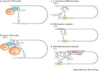
Several
groups of positive-strand RNA viruses containRNAs that are neither capped nor
polyadenylated. These viruses have evolved alternative strategies for
translation that use 3′-UTR enhancer elements to recruit eIF4F or eIFiso4F
and/or to base pair with their 5′-UTRs for loading
the
43S preinitiation complex at the 5′- start site. Satellite tobacco necrosis
virus (STNV – a positive strand RNA necrovirus) RNA contains within its
3′-UTR (just after the termination codon of the coat protein coding region) a
translational enhancer domain (TED) that promotes efficient cap-independent
translation when combined with the STNV 5′-UTR (for STNV-1 strain see Timmer etal.
1993). Although TED can functionally substitute for a 5′-cap structure, its
function in vitro is dependent on the presence of eIF4F and eIFiso4F
(Timmer et al. 1993). Indeed TED specifically binds eIF4E and eIF4iso4Ein
vitro (van Lipzig et al. 2002; Gazo et al. 2004), while the
5′-UTR of STNV-1 has the potential to base pair with TED and the 3′-end of 18S
rRNA (Timmer et al. 1993).
Thus
to promote cap-independent translation initiation, TED recruits the 43S
preinitiation complex by binding canonical cap-binding factors at the 3′-UTR,
while potential base pairing between the viral 5′- and 3′-UTRs would be
required for transfer of this 43S complex to the 5′-UTR to locate the
initiation codon. The existence of the 3′- to 5′-UTR pathway to recruit the
translational machinery is probably not unique to STNV TED, but is likely to
apply to other enhancer elements present within the 3′-UTRs of other
non-capped, non-polyadenylated virus RNAs. Barley yellow dwarf virus (BYDV – a
luteovirus) RNA also lacks a 5′-cap structure and poly (A) tail, but it harbors
a cap-independent BYDV translational element (BTE) functionally similar to TED
within the 3′-UTR (Wang et al. 1997; Guo et al. 2000). A BYDV
like BTE is present in all Luteoviruses, as well as in Dianthovirus [Red clover
necrotic mosaic virus (RCNMV), Mizumoto et al. 2003] and Necrovirus
[tobacco necrosis virus (TNV), Meulewaeter et al. 2004; Shen and Miller
2004], and contains the conserved sequence CGGAUCCUGGGAAACAGG, which also
functions when placed in the 5′-UTR. In its natural location this sequence has
the potential to base pair to the 5′-UTR (Wang et al. 1997; Guo et
al. 2000, 2001). BTE can recruit the translation machinery to the 3′-end
and deliver it to the 5′-UTR by a 3′–5′ RNA interaction (Wang et al.
1997).
The
delivery of the translational machinery to the 5′-end can occur due to
long-distance kissing-loop interactions between RNA hairpins in BTE and the
5′-UTR (Guo et al. 2001). Thus, TED and BTE behave in a similar way to
strongly stimulate cap-independent translation, without exhibiting any
conservation of sequence or secondary structure. Whether BTE or TED require
participation of canonical translation initiation factors for their action
remains to be investigated. Another distinct enhancer element identified in
Tombusvirus, Tomato bushy stunt virus (TBSV), has been termed the
3′-cap-independent translational enhancer. The TBSV 5′-UTR folds into a complex
RNA structure, which was recently demonstrated to physically interact with the
3′CITE in vitro (Fabian and White 2004). Formation of 5′–3′ RNA
interactions correlates well with efficient translation in vivo and
might support the transfer of the translational machinery from the 3′ to the
5′-end of the RNA as suggested for BTE and TED (Figure 5).

No comments:
Post a Comment